Multiple Sclerosis – Experimental Autoimmune Encephalomyelitis EAE Mouse Model
Investigate your compound’s efficacy on the inflammation and immune component of Multiple Sclerosis using the EAE mouse model
EAE Mouse Model Key Characteristics
Experimental autoimmune encephalomyelitis (EAE) mouse model is a relevant preclinical model to study efficacy of novel treatments against multiple sclerosis. The EAE mouse model replicates the key pathological and clinical features of human multiple sclerosis (MS), characterized by autoimmune-induced neuroinflammation, astrogliosis and axonal damage that manifest as motor, sensory, autonomic and cognitive disabilities.
EAE mouse model is established either by immunization (referred to as active EAE mouse model) or by T-cell transfer from mice immunized for active EAE (referred to as adoptive transfer EAE mouse model). Active EAE mouse model is induced by immunization with antigens such as myelin-oligodendrocyte protein (MOG), myelin basic protein (MBP) or proteolipid protein (PLP) together with Complete Freund’s adjuvant (CFA) accompanied by an intraperitoneal injection of pertussis toxin (PTX) on the day of immunization and two days later. Immunization via active EAE induction can also be set-up in rats. For the adoptive transfer model, donor mice are first immunized with CNS antigen, followed by splenocyte isolation with in vitro re-activation, and the transfer of encephalitogenic T-cells to acceptor mice.
✓ Peripheral inflammatory infiltrate (lymphocytes, macrophage) induced CNS inflammation, nerve injury, axon degeneration, and cell death.
✓ Progressive limb paralysis.
✓ Complementary PK/PD profiling services.

As a preclinical immunology CRO, InnoSer offers well-established and clinically relevant multiple sclerosis mouse models, complemented with standardized study protocols to ensure consistency and reproducibility of your results. In addition, InnoSer offers research services using the Cuprizone, and Lysolecithin mouse models. While the EAE mouse model allows you to focus on the inflammatory component of MS, the Cuprizone and Lysolecithin models enable you to test the efficacy of compounds directed at remyelination. However, as all models have their unique characteristics, we recommend discussing your study setup in close collaboration with our experts.
InnoSer’s research network comprises scientific experts working on multiple sclerosis, offering you the possibility to consult your compound’s MOA in-depth.
Example study timeline using MOG-induced EAE mouse model
The MOG (Myelin Oligodendrocyte Glycoprotein)-induced mouse model of Multiple Sclerosis (MS) represents one of the most well-established, chronic models of severe, monophasic, demyelinating encephalomyelitis belonging to the group of direct EAE mouse models induced by immunization. The MOG-induced EAE mouse model offers you with the possibility to perform both prophylactic (largest therapeutic window of all EAE models) and therapeutic studies, being equally suitable both for first-pass compound screen studies and disease efficacy studies.

Study timeline example in the MOG-induced EAE mouse model of MS. EAE is induced in 8-10-week-old C57BL/6J female mice by immunization (D0) with subcutaneous (SC) injection of MOG35-55 suspended in complete Freud’s adjuvant (CFA). One to two hours after (D0), mice are injected intraperitoneally (IP) with pertussis toxin (PTX). On D1, 24 hours after the first PTX injection, a second PTX injection is given to all mice again. In the MOG-induced EAE mouse model of MS, the MOG35-55 peptide initiates expansion and differentiation of MOG-specific autoimmune T cells, the PTX will further enhance disease development by modulating immunological response and facilitating the movement of autoimmune T cells into the CNS.
Prophylactic treatment will start on the day of immunization (D0) and will continue until study termination. Therapeutic treatment starts as each mouse develops EAE (D11-12) and depending on the compound’s characteristics will last until study termination (typically D28), and will include a randomization step to assign mice into groups based on their EAE scores
Motor function assessment using a rotatord (including –7D to –D3 training sessions, D9, D18, and D28) can be included as an option. Blood collection (D0, D18, and D28) can be included to assess plasma biomarkers (e.g., NfL) or to determine your compound’s PK/PD profile.
EAE Mouse Model Sample Data

MOG35-55 immunization in female 8-week-old C57BL/6J mice results in robust and severe acute monophasic, demyelination with peak disease (maximum EAE scores) onset 14-18 days after immunization.
In this study, we assessed the disease progression in the MOG-induced EAE mouse model and mice without EAE.

MOG35-55 immunization in female 8-week-old C57BL/6J mice results in progressive decrease in body weight correlating with neurological disease scoring.
Compared to uninduced mice, EAE-induced mice show a progressive decrease in body weight following MOG immunization throughout the course of the study. The initial drop in body weight of all groups was triggered by immune system reaction following PTX injection.

MOG35-55 immunization in female 8-week-old C57BL/6J mice results in progressive motor dysfunction as assessed by latency to fall on Rotarod correlating with neurological disease scoring.
Compared to uninduced mice, EAE-induced mice showed progressive decrease in latency to fall on Rotarod over the course of disease.

MOG₃₅–₅₅ immunization in female 8-week-old C57BL/6J mice induces neuroaxonal damage as assessed by increased plasma neurofilament light chain (NfL) levels measured by MSD.
Compared to non-induced (negative control) mice, vehicle-treated EAE mice showed a significant increase in plasma NfL at day 16 (**P<0.01) and 28 post-immunization (**P<0.01), corresponding to peak disease activity.
EAE Mouse Model Readouts
The People Behind Your Research

Hasselt University/BIOMED
As part of a joint initiative to advance preclinical MS research, InnoSer works together with researchers from BIOMED, who focus on immunological mechanisms, myelination, and damage processes in the brain during MS.
")
Céline Erens, PhD, Immunology Study Director
An expert team led by our immunology study director; Céline Erens works together with you to help you set up optimal study designs. Curating the preclinical testing of your lead compounds with a deep understanding of the field is your solution to accelerating your drug development.
Frequently Asked Questions
What is an EAE model?
The Experimental Autoimmune Encephalomyelitis (EAE) mouse model is a widely used in vivo model for studying the inflammatory and autoimmune pathophysiology of Multiple Sclerosis (MS). EAE mimics key aspects of MS pathology, including demyelination, neuroinflammation, and T-cell mediated autoimmunity. In mice, EAE is induced by immunization with myelin proteins or peptides, which triggers an autoimmune response targeting the central nervous system (CNS), leading to inflammation, demyelination, and neurological deficits similar to those seen in human course of MS.
Key features of the EAE mouse mode include:
- Pathophysiology: The model mimics the T-cell-mediated autoimmune response that causes demyelination and neuroinflammation in MS.
- Clinical signs: Symptoms include muscle weakness, paralysis, and gait abnormalities, which are scored to assess the disease progression.
At InnoSer, the EAE mouse model is widely used to evaluate the efficacy of immunomodulatory therapies, anti-inflammatory drugs, and novel therapies for MS treatment. Contact us to learn more about how InnoSer’s expertise using the EAE mouse model of MS can accelerate your preclinical research.
Is EAE a good preclinical model for efficacy studies of novel therapeutics for Multiple Sclerosis?
- T-cell-mediated mechanism: EAE involves an autoimmune attack on the central nervous system (CNS) similar to the immune response seen in MS, where T cells target myelin proteins.
- Demyelination and neuroinflammation: Like MS, EAE results in demyelination, neuroinflammation, and motor dysfunction, which can be used to evaluate the effects of anti-inflammatory and immunomodulatory treatments.
- Disease progression: EAE allows researchers to observe disease onset, progression, and clinical symptoms (such as paralysis and muscle weakness), providing insights into the mechanisms of MS.
Though EAE does not fully replicate all aspects of MS, particularly the chronic phase of the disease, it remains one of the most commonly used and validated models for MS research and drug development. In addition to the EAE mouse model, InnoSer offers research services using the Cuprizone, and Lysolecithin mouse models. While the EAE mouse model allows you to focus on the inflammatory component of MS, the Cuprizone and Lysolecithin models enable you to test the efficacy of compounds directed at remyelination.
However, as all models have their unique characteristics, we recommend discussing your study setup in close collaboration with our experts.
Reach out to our experts to discuss which mouse model of MS is the most suitable for your research.
Is there a difference in EAE onset and disease pattern dependent on the mouse strain you use?
- EAE-susceptible strains (e.g., C57BL/6) typically exhibit a Th1-type phenotype, characterized by the activation of pro-inflammatory T cells. These strains often show a more severe disease course, with faster onset and pronounced symptoms such as motor deficits and paralysis.
- EAE-resistant strains (e.g., BALB/c) generally have a Th2/Treg phenotype, which is associated with anti-inflammatory responses. These strains tend to exhibit a milder form of EAE, with delayed disease onset and less severe symptoms due to stronger regulatory T cell (Treg) activity and a greater production of anti-inflammatory cytokines.
The choice of mouse strain is crucial as it can influence the disease progression, immune cell response, and overall pathophysiology of the EAE model, making it essential for researchers to select the right strain based on their specific research objectives and the therapeutic mechanisms being studied. Reach out to our team to discuss and obtain guidance on selecting the optimal mouse strain for your EAE study.
Are there specific readouts you recommend running for preclinical efficacy studies in the EAE mouse model?
Selecting the right readouts for your EAE study is essential for assessing the efficacy of your novel immunomodulatory therapeutics. At InnoSer, we recommend a selection of following readouts to use in your EAE studies:
- Clinical scoring of symptoms such as paralysis, gait abnormalities, and muscle weakness is a critical readout. This helps track disease progression and response to treatment. A commonly used scale is the neurological score to quantify motor function.
- Motor function tests (e.g., rotarod, grip strength) can be used to quantitatively assess neurological impairment and functional recovery in response to therapeutic interventions.
- Measuring levels of cytokines (e.g., IFN-γ, TNF-α, IL-6, IL-10) helps you determine the Th1/Th2 balance and the nature of the immune response. Multiplex ELISA, meso-scale discovery (MSD), and/or flow cytometry can be further used for profiling cytokine secretion and immune cell activity in the EAE mouse model.
- Histopathology analyses performed by in-house veterinary pathologists allow you to assess the degree of inflammation and/or demyelination following treatment with your test compounds. This is essential for understanding the underlying mechanisms and validating the efficacy of your immunomodulatory compounds.
Discover Other Related Immunology Mouse Models


Stay Curious: More Articles to Explore
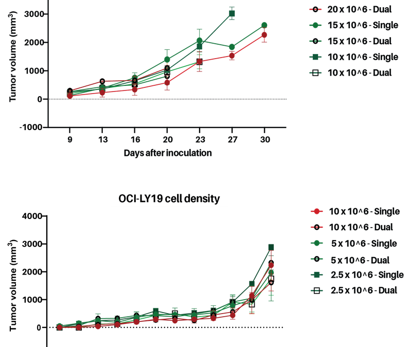
Personalized Lymphoma CDX mouse models for preclinical oncology
In recent studies, we established and validated two new human lymphoma cell‑line–derived xenograft (CDX) models, generated from the OCI‑Ly19 (diffuse large B‑cell lymphoma, DLBCL) and REC‑1 (mantle cell lymphoma) cell lines. These models were developed in strategic...

Multiplex Cytokine and Immune Cell Profiling in Immuno-Oncology Research
The Meso-Scale Discovery (MSD) platform is a high-sensitivity, multiplex cytokine profiling analysis system, enabling you to simultaneously quantify multiple target cytokines, chemokines, antibodies, and growth factors from minute sample volumes (as low as 1 µL). By...

Pancreatic Cancer PDX Models
Last month, November, represented Pancreatic Cancer Awareness Month; a time to focus on the challenges posed by one of the most aggressive and lethal cancer types. Pancreatic cancer accounts for approximately 3% of all diagnosed cancers in Europe, with pancreatic...

Exploring InnoSer’s Key In vitro Oncology Services: PD-1 PD-L1 Blockade Assay
Immune Checkpoint Blockade in Oncology Drug Discovery In recent years, immune checkpoint blockade has emerged as a groundbreaking therapeutic strategy in immuno-oncology. Programmed cell death 1 receptor (PD-1), and its ligand programmed cell death ligand 1 (PD-L1)...
AAALAC Accreditation
InnoSer has earned the AAALAC accreditation, demonstrating our commitment to responsible animal care and use. AAALAC International is a nonprofit organization that promotes the humane treatment of animals in science through voluntary accreditation and assessment programs. InnoSer’s facilities in the Netherlands and Belgium have been AAALAC-accredited since 2016 and 2020, respectively. Read more about the AAALAC accreditation programme here.
![]()
Animal Welfare
The 3Rs impact everything from policy and regulatory change to the development and uptake of new technologies and approaches. This is why InnoSer has ongoing commitment and monitoring of these processes. The steps we practice maximize our ability to replace, reduce and refine animal involvement and facilitate our commitment to these principles when it comes to research and drug development.
info@innoserlaboratories.com