Diabetes – Type 1 Diabetes Mouse Models
Test your novel investigational therapy’s effect on glycemic control and glucose tolerance thanks to InnoSer’s expertise in Type 1 diabetes mouse models
Type 1 Diabetes Mouse Models Key Characteristics
Type 1 Diabetes mouse models are established either via chemical induction (e.g., STZ or alloxan-induced diabetes) or via genetic modifications (e.g., NOD mouse model). STZ or alloxan chemically-induced models result in targeted beta cell destruction and are flexible for finetuning the level of beta cell ablation and the degree of hypoglycemia. The genetic NOD mouse model is prone to T1D development via autoimmune islet destruction, making them of particular interest to evaluate immune-modulatory drugs. Key readouts include glucose tolerance testing, evaluation of immune cell infiltration and islet destruction, beta cell and islet evaluation, blood biomarkers, hormone quantification, and histological and clinical endpoints.
InnoSer’s type 1 diabetes mouse models and readouts, therefore, arise as suitable efficacy models for therapies aimed to target auto-immunity, promote beta-cell regeneration, and facilitate research of novel approaches ranging from (stem) cell therapies, small molecules to hormone modulating approaches or gene therapies. Test your novel investigational therapy’s efficacy on glycemic control and glucose tolerance in a fast and golden standard experimental diabetes mouse model with full tailoring flexibility.
✓ Chemically induced (Alloxan, STZ) type 1 diabetes in mice.
✓ Study comorbidities such as diabetic kidney disease development.
✓ Suitable for assessment of novel ATMPs such as cell therapy or other regenerative strategies.
✓ Complementary in vitro immunoassays, histopathology analyses, and PK/PD profiling services.

Developing new, safe, and efficacious therapies is an extremely intricate process. As a preclinical immunology contract research organization (CRO), InnoSer partners with you to help you navigate the complexities of this research area.
Take advantage of InnoSer’s collaborative approach to develop the most optimal study design. With flexible and fast study start times you can perform your research at an accelerated pace. By outsourcing your preclinical oncology studies to InnoSer, you gain access to our in vitro and in vivo immunology drug development portfolio.
Type 1 Diabetes Mouse Models Sample Data

Early Signs of Diabetic Kidney Disease Following Alloxan-Induced T1D in Mice: Morphological and Biochemical Changes
Following chemical T1D diabetes induction (Alloxan), signs of development of early diabetic kidney disease such as distorted and enlarged glomeruli become apparent (B) compared to normal healthy (control) conditions (A). Quantitative analysis of the glomerulus area (in pixels) was performed in control mice and alloxan-induced diabetic mice. The diabetic group exhibited a significant increase in glomerulus area indicating glomerular hypertrophy, a characteristic feature of diabetic nephropathy(C). (D) BUN levels were significantly elevated in the diabetic group compared to healthy controls, indicating the development of diabetic kidney disease.

Islet Atrophy and Reduced Plasma C-Peptide Levels Following Alloxan-Induced T1D in Mice
Following chemical T1D diabetes induction (Alloxan), compared to control mice (A) type 1 diabetic mice show islet atrophy (B). Drop in mouse c-peptide plasma as a surrogate marker for insulin is observed in alloxan-type 1 induced mice compared to control mice.

Imaging of Transplanted Islets in Diabetic Mouse Model
(A) brightfield microscopy of transplanted islets under the kidney capsule in a diabetic mouse model. (B) immunofluorescent staining of the same islets, with pancreatic beta cells stained for insulin (green) and pancreatic alpha cells stained for glucagon (red). Nuclei are counterstained with DAPI (blue).

Glucose measurement for chemically induced and insulin treated diabetic mice after 14 days
Examine in vivo functionality of transplanted islet cell preparation after transplantation under the kidney capsule or subcutaneous transplantation sites. C-peptide levels are followed up to discriminate between insulin secreted by the mouse pancreas islets and transplanted cells. Diabetes reversal can be used to show therapeutic efficacy of your cell therapy product

Static Glucose Stimulated Insulin Secretion (GSIS) Assay
Static GSIS assays can be performed to examine the effects on insulin secretion after glucose stimulation. The assay can be performed with cell lines (INS1E, MIN6) or primary cell preparations.
Type 1 Diabetes Mouse Models Readouts
The People Behind Your Research

Yanick Fanton, PhD, Chief Scientific Officer
As Chief Science Officer at InnoSer, Yanick is responsible for all customer studies at InnoSer and takes care of the scientific and technical coordination.
")
Céline Erens, PhD, Immunology Study Director
An expert team led by our immunology study director; Céline Erens works together with you to help you set up optimal study designs. Curating the preclinical testing of your lead compounds with a deep understanding of the field is your solution to accelerating your drug development.

Leuven Diabetes Lab
InnoSer works closely with the lab of Prof. Dr. Chantal Mathieu, integrating their state-of-the-art type 1 diabetes expertise to help our clients design robust, translationally relevant preclinical studies, ultimately accelerating the development of new therapies.
Discover Other Related Immunology Mouse Models


Discover Other Related Metabolic Disease Mouse Models


Stay Curious: More Articles to Explore
Personalized Lymphoma CDX mouse models for preclinical oncology
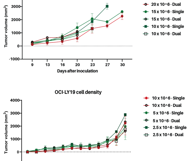
In recent studies, we established and validated two new human lymphoma cell‑line–derived xenograft (CDX) models, generated from the OCI‑Ly19 (diffuse large B‑cell lymphoma, DLBCL) and REC‑1 (mantle cell lymphoma) cell lines. These models were developed in strategic...

Multiplex Cytokine and Immune Cell Profiling in Immuno-Oncology Research
The Meso-Scale Discovery (MSD) platform is a high-sensitivity, multiplex cytokine profiling analysis system, enabling you to simultaneously quantify multiple target cytokines, chemokines, antibodies, and growth factors from minute sample volumes (as low as 1 µL). By...

Pancreatic Cancer PDX Models
Last month, November, represented Pancreatic Cancer Awareness Month; a time to focus on the challenges posed by one of the most aggressive and lethal cancer types. Pancreatic cancer accounts for approximately 3% of all diagnosed cancers in Europe, with pancreatic...

Exploring InnoSer’s Key In vitro Oncology Services: PD-1 PD-L1 Blockade Assay
Immune Checkpoint Blockade in Oncology Drug Discovery In recent years, immune checkpoint blockade has emerged as a groundbreaking therapeutic strategy in immuno-oncology. Programmed cell death 1 receptor (PD-1), and its ligand programmed cell death ligand 1 (PD-L1)...
AAALAC Accreditation
InnoSer has earned the AAALAC accreditation, demonstrating our commitment to responsible animal care and use. AAALAC International is a nonprofit organization that promotes the humane treatment of animals in science through voluntary accreditation and assessment programs. InnoSer’s facilities in the Netherlands and Belgium have been AAALAC-accredited since 2016 and 2020, respectively. Read more about the AAALAC accreditation programme here.
![]()
Animal Welfare
The 3Rs impact everything from policy and regulatory change to the development and uptake of new technologies and approaches. This is why InnoSer has ongoing commitment and monitoring of these processes. The steps we practice maximize our ability to replace, reduce and refine animal involvement and facilitate our commitment to these principles when it comes to research and drug development.
info@innoserlaboratories.com