
Tauopathy Mouse Models – Seeding and Spreading Tau mouse models
Accelerate decision-making with a Tau seeding and spreading mouse models that combine established Tau pathology with reproducible, prion-like spreading across connected brain regions
Tau Seeding Mouse Models Key Characteristics
Seeding tau mouse models represent a powerful preclinical tool for performing proof-of-concept and/or target engagement studies targeting primary tauopathies (frontotemporal dementia, Pick’s disease, corticobasal degeneration, progressive supranuclear palsy etc.,) as well as secondary tauopathies like Alzheimer’s disease.
In traditionally used transgenic models that overexpress Tau, pathology arises cell-autonomously in many neurons at the same time, in turn making it difficult to distinguish between intracellular development of tau pathology from true prion-like spreading of tau pathology across connected brain regions.
In mouse models of tau seeding and spreading, tau aggregates are injected into the hippocampus of transgenic tau mouse models. Tau aggregates are injected into transgenic tau mice at young ages whereby the mice show minimal or no tau aggregation pathology. In this way, endogenous (non-aggregated) tau in transgenic mice is used as a background, allowing controlled recruitment of tau aggregation and pathological propagation of the injected tau aggregates.
For Tau seeding studies, InnoSer works with Tau aggregates coming from various sources, including recombinant aggregates, human brain-derived extracts (Alzheimer’s. Disease or frontotemportal dementia-Tau) as well as aged transgenic Tau mouse brain extracts, injected into any transgenic line to study compound’s effects on the pathologic spread of Tau.
Therefore, if your therapeutic’s mechanism of action is aimed at inhibiting Tau propagation and pathological spread, tau seeding mouse models may be a more well-suited, translationally relevant alternative to transgenic models, allowing you to assess target engagement and early efficacy.
✓ InnoSer’s Transgenic homozygous Tau[P301S] mouse model injected with brain homogenates from homozygous aged Tau[P301S] mice shows strong seeding and spreading pattern over 9 weeks, enabling you to perform short-term proof-of-concept and/or target engagement and biomarker development studies
✓ In contrast to homozygous Tau[P301S], InnoSer’s heterozgyous Tau[P301S] seeding model shows lower seeding and spreading patterns, with astrocytosis (GFAP) and microgliosis (CD45), allowing you to evaluate neuroinflammation-related targets and endpoints
✓ To mimic disease pathophysiology more accurately, InnoSer also works with optimized Tau seeding mouse models using human Alzheimer’s disease brain extracts using transgenic Tau models such as the Tau[P301S] PS19 line and/or humanized Tau model (originally described by Andorfer et al., 2003)

Take advantage of InnoSer’s preclinical research expertise in modeling Tau pathology, flexibility, and collaborative approach for your research. Our in-house neurology experts have long-standing experience with modeling Tau pathology in vivo and are happy to help guide your decision on study design fit for your current research goals.
InnoSer’s neurology expert team possesses relevant experience in working with multiple therapy types, including small molecules, peptides, enzymes, oligonucleotides, gene therapy (viral vectors, e.g., AAVs), and immunotherapies (antibody/vaccine immunotherapies).
Your Alzheimer’s Disease Research Starts Here.
Explore our expertly curated comparison of available mouse models to make faster, data-driven decisions. View example study timeline, recommended readouts, and example data featuring validation datasets across the different mouse models.

Tau Seeding Mouse Models Sample Data

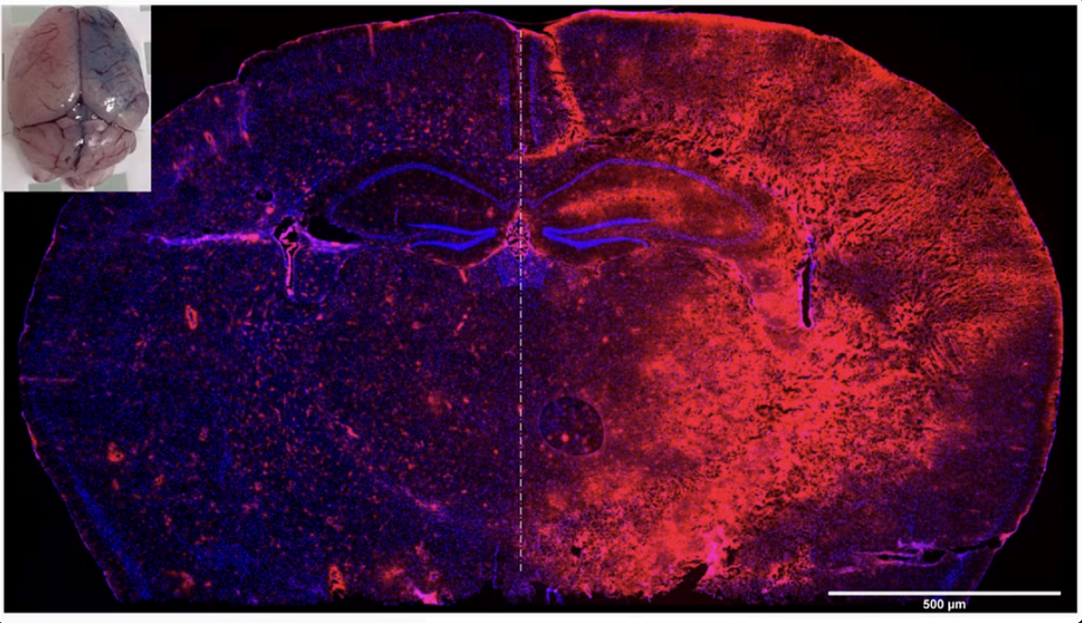
Seeding and spreading of Tau pathology (AT8) in homozygous Tau[P301S] mice injected with brain lysates from end-stage homozygous Tau[P301S] mice
Young (2-month-old) homozygous Tau[P301S] mice were injected in the CA1 region of the hippocampus with brain homogenate from homozygous aged Tau[P301S] mice (n=8) or with negative control (n=5).
Aggregated Tau was detected via IHC 9 weeks post-injection, showing robust seeding at the (A-B) injection site and (C-D) extensive spreading to the contralateral hippocampus.
(E) Significant correlation between pathological burden in the ipsilateral and contralateral hippocampus confirms efficient prion-like propagation of pathology in a human Tau-expressing background (Ahmed et al., 2014).
Quantification was performed on five sections per mouse (mean ± SEM). Statistics: Student’s t-test (* p < 0,05).
Tau Seeding Mouse Models Readouts
Don’t See Your Model? We Can Help!
Our experts specialize in tailored solutions to meet your unique research needs. Let’s discuss how we can support your project.
Preclinical Alzheimer’s disease Research with InnoSer

Validated Alzheimer’s disease mouse models
Choosing the right Alzheimer’s disease mouse model determines which pathological endpoints are measurable, which behavioral and biomarker readouts are appropriate, and how your data will be interpreted in a clinical context. Leverage our transgenic amyloid, tau, combined amyloid/tau, and tau seeding models. Read more >>

Translationally relevant Alzheimer’s disease biomarkers
Biomarkers are now central to Alzheimer’s disease clinical trials; generating translationally relevant preclinical readouts has never been more critical. Learn how you can implement clinically relevant biomarkers in one of our validated Alzheimer’s disease models, from plasma Aβ42/40 and/or CSF pan-Tau to the neurodegeneration marker, NfL. Read more >>

Functional behavioral endpoints
Behavioral and functional endpoints are how you demonstrate that your compound is reaching and rescuing cells in the brain regions relevant to Alzheimer’s disease. Assess cognitive and motor functions that have been validated across our Alzheimer’s disease models using CognitionWall™, Morris Water Maze, Rotarod, CatWalk, and ex vivo LTP. Read more >>
The People Behind Your Research

Sofie Carmans, PhD
Principal Scientist Neurology

Thomas Vogels, PhD
Principal Scientist Neurology
Struggling with getting your therapeutics across the blood-brain-barrier?
Discover how SonoCloud® ultrasound-mediated BBB disruption can improve brain uptake — without altering your compound.
Frequently Asked Questions
What are the advantages of choosing to work with a seeding and spreading tau model over a transgenic tau model?
InnoSer’s tau seeding and spreading mouse models offer many significant advantages over classical transgenic tau overexpression models, particularly when replication of pathological tau propagation and translational relevance are central to your therapeutic strategy and your compound’s mechanism of action (MoA).
Namely, therapies targeting tau propagation/intracellular spread, lysosomal or autophagy-enhancing strategies, as well as therapies addressing secondary disease processes across primary and secondary tauopathies like Alzheimer’s disease, may be more suitable for evaluation in seeding and spreading models. Given that transgenic mouse models are more aggressive in comparison to seeding models, they may be more suitable for direct disease targeting mechanism approaches (i.e., small molecules targeting aggregate pathways, direct therapies such as antibody approaches, RNA approaches – ASOs, siRNA, mRNA).
Compared to traditional single transgenic tau models, such as the Tau[P301S] models, including the PS19 line and/or the Tau[P301L] model, seeding and spreading tau mouse models allow you to perform proof-of-concept and/or efficacy studies in a more translationally relevant disease setting. Seeding and spreading tau models show rapid and robust tau pathology, including spreading and progression in a time-dependent manner that is frequently observed in human tauopathies. Compared to classical transgenic mouse models, where tau pathology occurs cell-autonomously, seeding and spreading mouse models feature prion-like tau propagation, recapitulating key features of human tauopathies, including intracellular spread, anatomical spread, and trans-synaptic propagation (Ahmed et al., 2017).
Importantly, the utilized tau seeds to induce pathology do not only have to be sourced from aged transgenic tau models or synthetic aggregates, but also from defined patient populations, enabling evaluation of strain-specific pathology and increasing translational relevance for precision medicine approaches in Alzheimer’s disease, FTD, or other primary and secondary tauopathies.
Tau seeding models typically allow shorter and more controlled study durations compared to aging-based transgenic models. Because pathology is induced at a defined time point, you can benefit from predictable and quick progression windows (e.g., in our Tau[P301S] seeding and spreading studies, the study endpoint is typically 9 weeks post-injection).
Ultimately, these characteristics make it a highly flexible platform for the development and testing of tau-based treatment strategies.
Is there a species barrier when using human patient-derived tau aggregates in InnoSer’s tau seeding and spreading mouse models?
A species barrier restricts prion transmissions across species, determining the infectivity of pathogenic prions. Given that tauopathies present in a similar way to prion diseases, working with human patient-derived tau aggregates injected into mouse models could imply that pathogenic human tau aggregates would not induce pathology due to “incompatibility” between the two tau species – from human and mouse origins, respectively.
In InnoSer’s Tau seeding and spreading models, this is not a limiting factor, as we use transgenic mice expressing human tau. Although low levels of endogenous mouse tau will be present in these transgenic tau mouse models, they will not act as the main substrate for aggregation in models of seeding and spreading.
Similarly, at the ages (2 months of age) for tau seeding experiments, endogenous pathological tau is minimal, and the injected human tau aggregates interact primarily with soluble (non-aggregated) human tau expressed by the transgene of the mouse model, enabling efficient prion-like propagation of tau in accordance with the scientific literature (Ahmed et al., 2014; Clavaguera et al.,2009).
This design allows robust and reproducible assessment of Tau seeding, spreading, and target engagement in a human-relevant Tau context.
Can InnoSer work with patient-derived material for Tau seeding studies?
Yes, as a preclinical CRO specialized in neurodegenerative disease models and tauopathies, InnoSer frequently works with patient-derived tau material for in vivo tau seeding studies. InnoSer has access to post-mortem brain extracts obtained from patients with tau-relevant neurodegenerative diseases, including frontotemporal dementia (FTD), Alzheimer’s disease, and other primary and secondary tauopathies.
In addition to working with patient-derived material, InnoSer also performs seeding studies using aged brain extracts from the Tau[P301S] mouse model, recombinant Tau fibrils (Stressmarq) and/or synthetic preformed fibrils (PFFs).
While InnoSer frequently performs studies using in-house established and validated seeding mouse models, including Tau[P301S] using seeds from end-stage mice of the same line, and/or the Tau[P301L] using recombinant seeds, we have also carried out multiple studies using human brain extract samples from multiple tauopathies (including Alzheimer’s disease and frontotemporal dementia) in commonly utilized tau lines such as the PS19 and/or the hTau model (described by Andorfer et al.,2003).
Where are the tau aggregates injected, and to which brain regions does it spread?
In InnoSer’s tau seeding mouse models, tau aggregates are delivered via stereotactic injection into the hippocampus, typically targeting the CA1 region. Following injection, we observe rapid induction of Tau pathology locally (i.e., 9 weeks post-injection in the Tau[P301S] mouse model; you can refer to a subset of the validation data here), with subsequent anatomically connected spread across the hippocampal formation, including the dentate gyrus and CA fields, as well as propagation toward the entorhinal cortex and cortical regions. Further statistical analyses (correlation of pathology, AT8+ signal, in the two regions) performed by InnoSer’s team suggest that the observed distribution pattern reflects pathological spread from the ipsilateral to contralateral sides rather than diffusion of tau aggregates from the injection itself.
InnoSer’s robust in-house characterization of induction kinetics, anatomical progression, including semi-quantitative data on tau pathology, as well as related neuroinflammation to ensure high scientific validity and reproducibility for preclinical anti-Tau therapeutic testing.
Interested in reviewing our full validation dataset or discussing whether a Tau seeding model fits your mechanism of action?
Reach out to InnoSer’s experts to obtain full validation slide decks.
Difference between heterozygous and homozygous Tau[P301S] seeding mouse model?
InnoSer’s tau[P301S] mouse model arises as a highly suitable background mouse model to study efficacy of therapeutics on pathologic tau seeding and propagation. Both heterozygous and homozygous Tau[P301S] genotypes are available for studies, with each differing in resulting pathology.
In InnoSer’s studies, both genotypes are typically injected with aged brain extracts obtained from end-stage (5-5.5 months old) homozygous Tau[P301S] mice due to the fact that the overexpression of human mutant P301S tau is more aggregation-prone than other mutations.
Due to higher transgene expression, homozygous mice develop faster and more aggressive Tau pathology. Typically injected at ~2 months of age, they show robust seeded pathology within 9 weeks post-injection, making them highly suitable for rapid proof-of-concept and direct anti-Tau efficacy studies.
In contrast, heterozygous Tau[P301S] mice exhibit lower levels of Tau seeding and spreading. When injected at ~6 months of age and analyzed 9 weeks later, they develop measurable Tau pathology accompanied by astrogliosis and microgliosis, providing a more moderate and extended window to evaluate therapeutic effects on both Tau propagation and associated neuroinflammation.
InnoSer’s Available Alzheimer’s Disease Model Types

Amyloid (APP/ AB) Transgenic Mouse Models
InnoSer offers preclinical research services with several different transgenic amyloid models, which recapitulate the plaque pathology of AD.

Transgenic Tau Mouse Models
InnoSer offers unique research services with several different transgenic tau models, which recapitulate the Tau neurofibrillary tangle pathology of AD.

Tau Seeding & Spreading Mouse Models
InnoSer uses an AD brain extract injection model, providing unique preclinical services with a translational model of Tau pathology seeding and spreading.

In Vitro Neurology Assays
Screen your lead candidate compounds using InnoSer’s in vitro neurology assays to progress to preclinical in vivo studies with confidence
InnoSer’s Available Alzheimer’s Disease Mouse Models

Transgenic PS19 Mouse Model
Leverage one of the most widely used mouse models in preclinical research to evaluate the efficacy of your compound targeting tau pathology
![APP[V717I] mouse model](https://www.innoserlaboratories.com/wp-content/uploads/2026/05/APPV717I-mouse-model.png)
APP[V717I] mouse model

Tau[P301S] Mouse Model
Leverage InnoSer’s proprietary Tau[P301S] mouse model with reproducible and aggressive Tau pathology for fast, decision-driven preclinical efficacy studies
![APP[V717I] x PS1[A246E] mouse model](https://www.innoserlaboratories.com/wp-content/uploads/2026/05/APPV717I-x-PS1A246E-mouse-model.png)
APP[V717I] x PS1[A246E] mouse model
Test the efficacy of therapies targeting amyloid-beta accumulation, neuroinflammation, and cognitive impairment in an early-onset amyloidosis transgenic APPxPS1 Alzheimer’s disease model
![Tau[P301L] Mouse Model](https://www.innoserlaboratories.com/wp-content/uploads/2026/05/TauP301L-Mouse-Model.png)
Tau[P301L] Mouse Model
Leverage InnoSer’s Tau[P301L] mouse model with progressive, well-characterized Tau pathology for mechanism-driven preclinical efficacy studies

Transgenic APP x PS1 ARTE10 mouse model
Advance your amyloid-lowering therapeutic program by leveraging the widespread amyloid-beta pathology of the ARTE10 mouse model for robust preclinical efficacy studies
![APP[V717I] x Tau[P301S] mouse model, european neurology CRO specialists](https://www.innoserlaboratories.com/wp-content/uploads/2026/05/APPV717I-x-TauP301S-mouse-model.png)
APP[V717I] x Tau[P301S] mouse model
Evaluate multi-target therapeutics in InnoSer’s combined APPxTau disease model
Discover InnoSer’s Latest Research
Neurodevelopmental disorder mouse models at InnoSer — TSC, Rett & STXBP1
in this month's update, we invite you to explore recent developments in rare neurodevelopmental disorders associated with epilepsy and seizure phenotypes. We highlight emerging therapeutic approaches, discuss the importance of selecting translationally relevant...

Plasma NfL, p-tau217, or Aβ42/40: Which Alzheimer’s disease biomarkers should you include in your preclinical efficacy study?
A practical guide to preclinical Alzheimer’s disease biomarkers panel selection when you’re considering outsourcing to a specialist neurology CRO Biomarkers have transformed Alzheimer's disease clinical trials, enabling diagnosis confirmation for trial eligibility,...

Transgenic Amyloid, Tau, Seeding, or combined Amyloid/Tau pathology mouse models — which is the right one for your drug development program?
A practical guide to model selection when outsourcing your Alzheimer's disease efficacy study to a specialist neurology CRO Choosing the right Alzheimer's disease mouse model is one of the most consequential early decisions in a preclinical drug development program....

Morris Water Maze, Rotarod and beyond: How do you build a comprehensive behavioral test battery when outsourcing your preclinical Alzheimer’s disease efficacy study?
A practical introduction to behavioural test battery selection when outsourcing your Alzheimer's disease efficacy study to a specialist neurology CRO When outsourcing a preclinical Alzheimer's disease efficacy study, behavioral endpoint selection is one of the most...
AAALAC Accreditation
InnoSer has earned the AAALAC accreditation, demonstrating our commitment to responsible animal care and use. AAALAC International is a nonprofit organization that promotes the humane treatment of animals in science through voluntary accreditation and assessment programs. InnoSer’s facilities in the Netherlands and Belgium have been AAALAC-accredited since 2016 and 2020, respectively. Read more about the AAALAC accreditation programme here.

Animal Welfare
The 3Rs impact everything from policy and regulatory change to the development and uptake of new technologies and approaches. This is why InnoSer has ongoing commitment and monitoring of these processes. The steps we practice maximize our ability to replace, reduce and refine animal involvement and facilitate our commitment to these principles when it comes to research and drug development.
info@innoserlaboratories.com