Double Transgenic APPxPS1 Mouse Models – APP[V717I]xPS1[A246E] mouse model
Test the efficacy of therapies targeting amyloid-beta accumulation, neuroinflammation, and cognitive impairment in an early-onset amyloidosis transgenic APPxPS1 Alzheimer’s disease model
Characteristics of the transgenic APPxPS1 mouse model of Alzheimer’s disease
The double transgenic APP[V717I]xPS1[A246E] mouse model represents a mouse model commonly used to perform preclinical efficacy studies of novel Alzheimer’s disease therapeutics. This APPxPS1 model harbors two familial Alzheimer’s disease–associated mutations in the human amyloid precursor protein (APP[V717I]) and presenilin-1 (PS1[A246E]) genes, both governed under the murine Thy-1 promoter. Clinical mutations in APP and presenilins are known causes of early-onset familial Alzheimer’s disease, driving increased amyloid-β production and altered Aβ42/Aβ40 ratios.
Originally described by Dewachter et al. (2000), this double transgenic APP×PS1 model was developed as a more aggressive complement to late-onset single transgenic APP line featuring the “London” ([V717I]) mutation associated with familial Alzheimer’s disease (FAD). Indeed, compared to the APP[V717I] single transgenic mice, the APPxPS1 model demonstrates accelerated amyloid pathology, earlier plaque onset, and a more pronounced amyloid burden. This makes it particularly suitable for evaluating amyloid-lowering therapies, disease-modifying strategies, neuroinflammatory mechanisms, and cognition-related endpoints within a shorter experimental timeline.
Belonging to a broader class of APPxPS1 transgenic mouse models which combine mutant human APP and PS1 or PS2 to model cerebral beta amyloidosis, these models are widely used in preclinical efficacy studies of amyloid-lowering therapies. In addition to performing efficacy studies in the APP[V717I]xPS1[A246E] line, InnoSer frequently performs efficacy studies in widely available APPxPS1 lines, including the ARTE10 mouse model of Alzheimer’s disease. Although both models display robust amyloid pathology, important differences exist in pathology onset, disease progression, phenotype and optimal study timelines. These distinctions are critical when selecting the most suitable model for your therapeutic program and are outlined in detail in our comparative FAQs.
✓ APPxPS1 mice show progressive β-amyloid plaque development in cortex, hippocampus and subiculum from an age of 6 months onwards, concomitant with development amyloid-associated neuroinflammation (microgliosis and astrocytosis)
✓ APPxPS1 mice show cognitive impairment in the Morris water maze paradigm and hippocampal LTP deficit at 8 months (not tested earlier)
✓ Pyroglutamate-modified Aβ42 (Aβ3(pE)-42) is detected in the insoluble brain fraction in APPxPS1 mice from 7 months onwards
✓ APPxPS1 mice show CAA pathology and micro-bleedings from 8 and 12-15 months of age, respectively, relevant for evaluation of amyloid-related imaging abnormalities (ARIA) in preclinical research

Take advantage of InnoSer’s expertise, flexibility, and collaborative approach for your research. We support you in identifying new drug candidates, characterizing their pharmacological properties, and conducting rigorous safety and efficacy studies with state-of-the-art behavioral, bioanalytical, and histopathological readouts.
Example data featuring the combined APP[V7I7I]xTau[P301S] mouse model
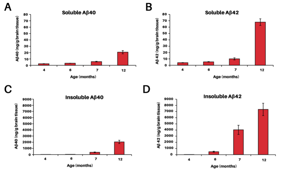
APP[V717I]xPS1[A246E] mice show progressive increase of (in)soluble Aβ 40/42 fibrils in cortex
Cortical levels of soluble (A) Aβ40 and (B) Aβ42, and insoluble (C) Aβ40 and (D) Aβ42, were quantified using ELISAs specific for human Aβ peptides (Thermo Fisher, KHB3481 & KHB3544). An age-dependent increase in both soluble and insoluble Aβ species was observed (N = 8–10 per group), demonstrating progressive amyloid pathology in this transgenic model.
Total (anti-Aβ nanobody) and dense plaque load (Thioflavin S) in subiculum of APP[V717I]xPS1[A246E] transgenic mice
(A) Quantification of total plaque load using anti-Aβ nanobody shows an age-dependent increase in amyloid deposition. (B) Representative IHC images corresponding to total plaque load. (C) Quantification of dense-core plaques using Thioflavin S staining also reveals progressive plaque accumulation. (D) Representative Thioflavin S-stained IHC images. N = 10 per group.
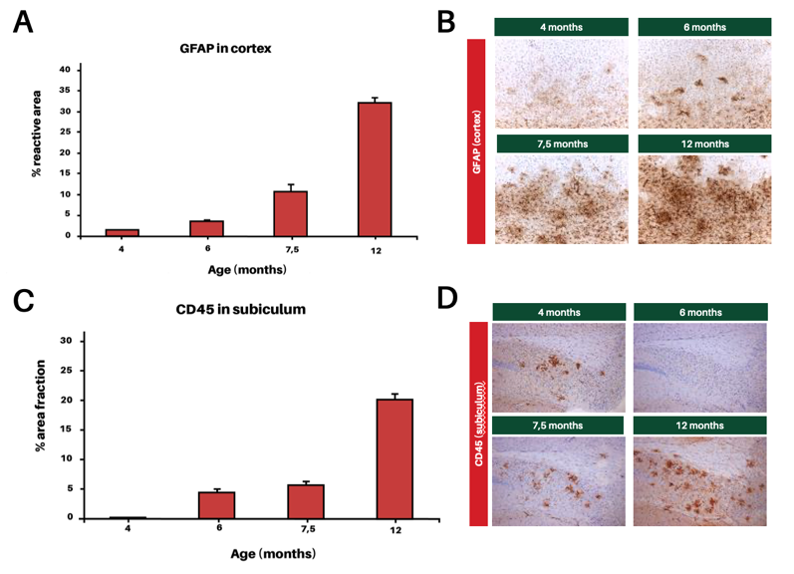
Brain inflammation (GFAP, CD45) in APP[V717I]xPS1[A246E] mice
(A) Quantification of astrocytosis using GFAP IHC and (B) representative images demonstrate increased astrocytic activation with age. (C) Quantification of microgliosis using CD45 immunohistochemistry and (D) representative IHC images show a parallel increase in microglial activation. N = 5–9 per group.
Your Alzheimer’s Disease Research Starts Here.
Explore our expertly curated comparison of available mouse models to make faster, data-driven decisions. View example study timeline, recommended readouts, and example data featuring validation datasets across the different mouse models.

Key readouts in the double transgenic APPxPS1 mouse model of Alzheimer’s disease
The People Behind Your Research

Sofie Carmans, PhD
Principal Scientist Neurology

Thomas Vogels, PhD
Principal Scientist Neurology
Frequently Asked Questions
How does the InnoSer’s APP[V717I]xPS1[A246E] line differ from the APPxPS1 ARTE10 mouse line of Alzheimer’s disease?
InnoSer’s APP[V717I]xPS1[A246E] mouse model develops amyloid plaques in the subiculum, hippocampus, and cortex from approximately 6 months of age, accompanied by progressive accumulation of soluble and insoluble Aβ40 and Aβ42 and pronounced neuroinflammation (GFAP and microglial activation). Cerebral amyloid angiopathy (CAA) emerges relatively early (from ~8 months), with microbleeds reported at later stages (12–15 months), making this model particularly relevant for vascular amyloid pathology and safety pharmacology studies. Importantly, APPxPS1 mice show robust spatial reference memory deficits in the Morris Water (see also figures 1 and 2 of Easton et al., 2013). Lastly, InnoSer’s proprietary APPxPS1 model is maintained within an established in-house breeding colony, allowing precise age-controlled study initiation and predictable availability.
Similarly, the ARTE10 mouse model is characterized by early-onset dense-core plaques and a high maximal plaque burden, with progressive deposition of human Aβ from around 6 months of age. Compared to InnoSer’s APPxPS1, advanced cerebral amyloid angiopathy has been described at later ages in the ARTE10 line (around 19 months) (Willuweit et al., 2009). While ARTE10 mice exhibit amyloid-associated neuritic changes and neuroinflammation, full neurofibrillary tangle pathology is absent, similar to other amyloid-driven models.
While both double transgenic APP×PS1 lines are well suited for evaluating amyloid-lowering strategies, the optimal choice depends on your required plaque burden, vascular endpoints (CAA/ARIA), cognitive readouts, and study timelines.
How does the double transgenic APP[V717I]xPS1[A246E] mouse model compare to the single transgenic APP[V717I] mouse model?
Both the APP[V717I]xPS1[A246E] mouse model and the APP[V717I] mouse model are widely used in preclinical Alzheimer’s disease research and help you generate comparable preclinical efficacy readouts, including amyloid-β (Aβ) accumulation, plaque pathology, and downstream functional impairments. However, the key difference lies in disease kinetics and amyloid burden.
The double transgenic APP×PS1 model was developed as a more aggressive complement to the late-onset APP single transgenic line featuring the “London” mutation ([V717I]), associated with early onset of Alzheimer’s disease (Dewachter et al., 2000). This familial Alzheimer’s disease mutation increases total Aβ production and shifts processing toward the more aggregation-prone Aβ42 species, thereby promoting amyloid plaque formation in an age-dependent manner.
However, by introducing the human PS1[A246E] mutation in addition to APP[V717I], the resulting combined model features a more aggressive and accelerated amyloid pathology phenotype (Dewachter et al., 2000). The PS1[A246E] mutation is a clinically identified early-onset familial Alzheimer’s disease mutation located in the transmembrane domain of presenilin-1, a key component of the γ-secretase complex. Expression of mutant PS1 under the murine Thy-1 promoter further enhances γ-secretase–mediated cleavage toward Aβ42 production. As a result, the double transgenic mice show a marked increase in brain Aβ42 levels and a dramatic elevation of the Aβ42/Aβ40 ratio compared to APP single transgenic mice (Dewachter et al., 2000), translating into accelerated amyloid pathology.
While APP[V717I] mice develop plaques typically around 10 months of age, APP[V717I]×PS1[A246E] mice exhibit robust plaque deposition as early as 6 months. Plaques in the double transgenic model are predominantly Aβ42-rich, reflecting the strong biochemical impact of the PS1 mutation, compared to single APP mice which show increase in brain Aβ40 at 15 months of age.
Therefore, from a practical perspective, the APPxPS1 model provides you with a shorter and more aggressive amyloid timeline, enabling faster evaluation of amyloid-lowering therapies, disease-modifying strategies, and cognition-related endpoints. The APP[V717I] single transgenic model, in contrast, may be preferred when studying slower, age-dependent amyloid progression.
At what ages are amyloid beta plaques observed in InnoSer’s APPxPS1 mouse line?
In InnoSer’s APP[V717I]xPS1[A246E] mouse model, total amyloid-beta plaque and dense-core plaques (Thioflavin S+) accumulation in the subiculum can be detected from approximately 6 months of age with high levels of amyloid burden visible at 12 months of age, during which robust amyloid pathology is observed. Similarly, robust cortical Aβ40 and Aβ42 deposition is observed from 6 to 12 months of age and beyond.
In this mouse model, the amyloid pathology is accompanied by age-dependent neuroinflammation, including astrocytosis (GFAP) and microgliosis (CD45), as well as elevated levels of the neuronal injury biomarker neurofilament light (NfL) in CSF and plasma from around 9 months of age.
Does InnoSer’s APPxPS1 mouse model display tau pathology alongside B-amyloid pathology?
While APPxPS1 mice develop robust amyloid pathology and dystrophic neurites containing hyperphosphorylated murine tau, they do not recapitulate full neurofibrillary tangle pathology.
This absence of overt tangle pathology is consistent with other APP/PS1 transgenic mouse models. Amyloid-only models robustly reproduce cerebral beta amyloidosis but do not recapitulate the full spectrum of Alzheimer’s disease encompassing tau pathology. To model both amyloid plaques and neurofibrillary tangles in vivo, the incorporation of mutant human tau is required.
Therefore, for therapies targeting combined amyloid-and-tau disease modification, we recommend the APP[V717I]xTau[P301S] mouse model, which recapitulates both extracellular amyloid plaques and progressive tau pathology, providing a more complete Alzheimer’s disease phenotype.
Learn more about InnoSer’s combined amyloid and tau mouse model here.
Does the APPxPS1 mouse model show cognitive deficits?
Yes, APPxPS1 mice demonstrate robust impairments in spatial reference memory in the Morris Water Maze (MWM) task. During acquisition training, transgenic animals show delayed learning compared to controls. In probe trials, the transgenic APPxPS1 mice show significantly reduced spatial reference memory compared to non-transgenic controls (see also figures 1 and 2 of Easton et al., 2013).
As an alternative in the APPxPS1 model, synaptic and memory-related deficits can be evaluated using electrophysiological readouts using ex vivo brain slices, such as hippocampal long-term potentiation (LTP), which provide sensitive measures of synaptic plasticity that can serve as a proxy measure for memory deficits in APPxPS1 mice. Indeed, APPxPS1 mice exhibit reduced potentiation response compared to wild-type mice ex vivo (click here to view the data).
For programs where cognitive improvement is a primary endpoint, InnoSer’s APP[V717I]xPS1[A246E] mouse model may offer greater sensitivity, as this model demonstrates clear spatial memory deficits in the Morris water maze along with documented compound-mediated rescue effects (see also figures 1 and 2 of Easton et al., 2013).
Has disease modification been demonstrated in the APPxPS1 mouse model?
Yes, published research has shown that disease modification has been demonstrated in the APP[V717I] mouse model in preclinical studies evaluating the efficacy of acetylcholinesterase inhibitors (Easton et al., 2013), GLP-1 receptor agonist (Hansen et al., 2016) and anti-PD1 antibodies (Latta-Mahieu et al., 2017).
Data from a study (Easton et al., 2013) has shown a significant improvement in reference memory in APPxPS1 mice along with a dose-dependent reduction in brain Aβ. These results suggest that donepezil may alleviate cognitive impairments in Alzheimer’s disease, in part, by reducing brain Aβ.
Has cerebral amyloid angiopathy (CAA) been described in InnoSer’s APPxPS1 mouse model and why is it relevant?
Yes, at ≥8 months of age, APPxPS1 mice exhibit CAA, marked by deposition of Aβ in vessel walls. This vascular amyloid accumulation leads to progressive vessel wall damage, aneurysm formation, and ultimately cerebral microbleeds by 12–15 months, mirroring vascular amyloidosis observed in a subset of AD patients.
Additionally, pyroglutamate-modified Aβ₃(pE)-42, a pathogenic and aggregation-prone Aβ species found abundantly in human AD plaques, is detected in the insoluble brain fraction of APPxPS1 mice from 7 months onward.
Cerebral amyloid angiopathy (CAA) is a common and clinically relevant cerebrovascular pathology characterized by the accumulation of Aβ peptides within the walls of cerebral blood vessels. CAA is present in a substantial proportion of Alzheimer’s disease (AD) patients and is increasingly recognized as a key contributor to vascular dysfunction, impaired cerebral blood flow, blood–brain barrier disruption, and intracerebral haemorrhage.
In recent years, interest in CAA has grown markedly as clinical trial outcomes have highlighted vascular amyloid as a potential driver of treatment-related adverse events, including amyloid-related imaging abnormalities (ARIA). Consequently, CAA has emerged as an important target for mechanistic studies and for the preclinical evaluation of anti-amyloid therapies, particularly immunotherapies and approaches aimed at improving vascular amyloid clearance.
Is InnoSer’s APPxPS1 mouse model readily available for preclinical efficacy studies?
Yes, as a preclinical neurodegeneration CRO, InnoSer maintains access to established breeding cohorts of the APPxPS1 mouse model, enabling rapid study initiation depending on the required animal age and genotype.
Our proactive colony planning ensures that your preclinical efficacy studies can be launched with minimal lead time.
InnoSer’s Available Alzheimer’s Disease Model Types
Amyloid (APP/ AB) Transgenic Mouse Models
InnoSer offers preclinical research services with several different transgenic amyloid models, which recapitulate the plaque pathology of AD.

Transgenic Tau Mouse Models
InnoSer offers unique research services with several different transgenic tau models, which recapitulate the Tau neurofibrillary tangle pathology of AD.

Tau Seeding & Spreading Mouse Models
InnoSer uses an AD brain extract injection model, providing unique preclinical services with a translational model of Tau pathology seeding and spreading.

In Vitro Neurology Assays
Screen your lead candidate compounds using InnoSer’s in vitro neurology assays to progress to preclinical in vivo studies with confidence
InnoSer’s Available Alzheimer’s Disease Mouse Models

Transgenic PS19 Mouse Model
Leverage one of the most widely used mouse models in preclinical research to evaluate the efficacy of your compound targeting tau pathology
![APP[V717I] mouse model](https://www.innoserlaboratories.com/wp-content/uploads/2026/05/APPV717I-mouse-model.png)
APP[V717I] mouse model

Tau[P301S] Mouse Model
Leverage InnoSer’s proprietary Tau[P301S] mouse model with reproducible and aggressive Tau pathology for fast, decision-driven preclinical efficacy studies
![APP[V717I] x PS1[A246E] mouse model](https://www.innoserlaboratories.com/wp-content/uploads/2026/05/APPV717I-x-PS1A246E-mouse-model.png)
APP[V717I] x PS1[A246E] mouse model
Test the efficacy of therapies targeting amyloid-beta accumulation, neuroinflammation, and cognitive impairment in an early-onset amyloidosis transgenic APPxPS1 Alzheimer’s disease model
![Tau[P301L] Mouse Model](https://www.innoserlaboratories.com/wp-content/uploads/2026/05/TauP301L-Mouse-Model.png)
Tau[P301L] Mouse Model
Leverage InnoSer’s Tau[P301L] mouse model with progressive, well-characterized Tau pathology for mechanism-driven preclinical efficacy studies

Transgenic APP x PS1 ARTE10 mouse model
Advance your amyloid-lowering therapeutic program by leveraging the widespread amyloid-beta pathology of the ARTE10 mouse model for robust preclinical efficacy studies
![APP[V717I] x Tau[P301S] mouse model, european neurology CRO specialists](https://www.innoserlaboratories.com/wp-content/uploads/2026/05/APPV717I-x-TauP301S-mouse-model.png)
APP[V717I] x Tau[P301S] mouse model
Evaluate multi-target therapeutics in InnoSer’s combined APPxTau disease model
Discover InnoSer’s Latest Research
STXBP1 Foundation and InnoSer validate R122X, a patient-variant mouse model of STXBP1 encephalopathy, to help expand therapeutic opportunities in STXBP1 research
Gene therapies, antisense oligonucleotides (ASOs), RNA editing technologies, and other precision medicine approaches are rapidly transforming the therapeutic landscape for STXBP1 encephalopathy. Currently, around 16 candidate therapies are in development across five...
![Cognitive profiling in the APP[V717I]xTau[P301S] mouse model](https://www.innoserlaboratories.com/wp-content/uploads/2026/07/Figure-1-MWM.png)
Cognitive profiling in the APP[V717I]xTau[P301S] mouse model
In this month's update, we revisit the APP[V717I]xTau[P301S] model with newly in-house generated Morris Water Maze (MWM) data, reconfirming the spatial memory deficits previously described for this model and reinforcing the translational value of the model.The...
![Translational Neuroscience: Comprehensive longitudinal profiling of the Tau[P301S] female vs male mice](https://www.innoserlaboratories.com/wp-content/uploads/2026/06/Female-TauP301S-mice-show-early-spontaneous-hyperactivity-in-automated-home-cages-PhenoTyperTM-229375_1080x323.png)
Translational Neuroscience: Comprehensive longitudinal profiling of the Tau[P301S] female vs male mice
In this month’s newsletter, we highlight and build upon our knowledge working with the Tau[P301S] mouse model (originally described by Allen et al., 2002), and its applicability in preclinical research.Female and male...

Clinically relevant biomarker panels for comprehensive efficacy studies in Alzheimer’s Disease mouse models
InnoSer's validated Alzheimer's disease (AD) mouse models are equipped with translationally relevant biomarker readout panels — spanning amyloid-beta (Aβ) species, phosphorylated Tau isoforms, and the early, sensitive hallmark of neurodegeneration, neurofilament light...
AAALAC Accreditation
InnoSer has earned the AAALAC accreditation, demonstrating our commitment to responsible animal care and use. AAALAC International is a nonprofit organization that promotes the humane treatment of animals in science through voluntary accreditation and assessment programs. InnoSer’s facilities in the Netherlands and Belgium have been AAALAC-accredited since 2016 and 2020, respectively. Read more about the AAALAC accreditation programme here.
![]()
Animal Welfare
The 3Rs impact everything from policy and regulatory change to the development and uptake of new technologies and approaches. This is why InnoSer has ongoing commitment and monitoring of these processes. The steps we practice maximize our ability to replace, reduce and refine animal involvement and facilitate our commitment to these principles when it comes to research and drug development.
info@innoserlaboratories.com